
See WhatWe See
Most people have never really seen their own teeth. A mirror shows you the front of them. It does not show you what is between them, what is under an old filling, or what is happening below the gum.
The equipment here does, and the screen faces you. When Dr. Cortes says a tooth needs work, you are looking at the same picture he is, at the same time, and you decide from there.
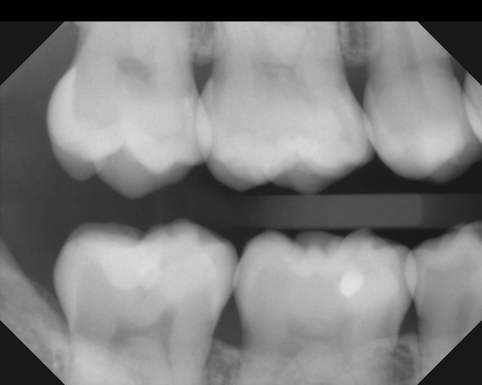
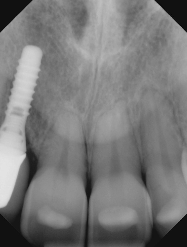
DigitalX-rays
Digital sensors replaced film years ago, and for good reason. The image is on screen in seconds rather than minutes, it can be enlarged and adjusted so small changes are visible, and the radiation dose is a fraction of what film required. It also means your images travel with your record instead of living in an envelope.
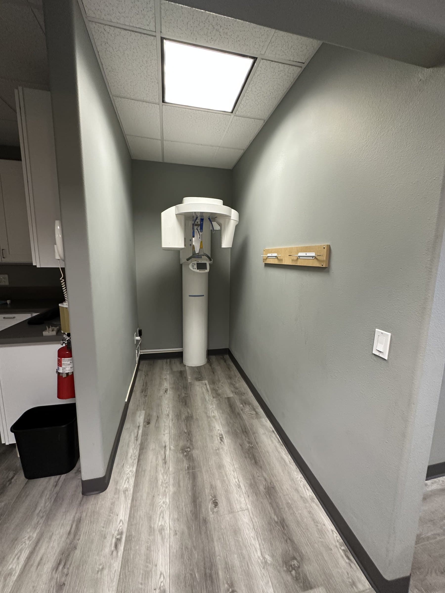
3D Imaging,CBCT
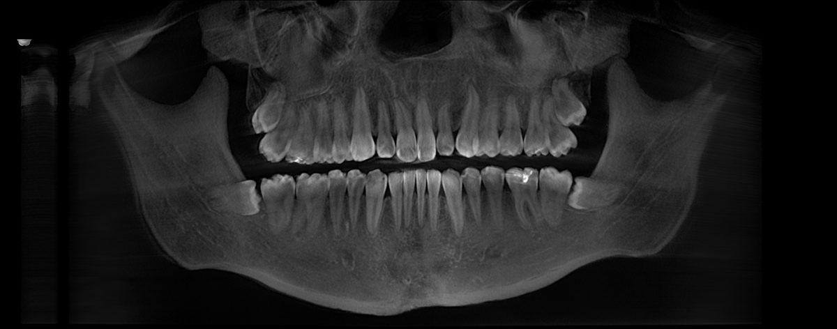
A standard X-ray flattens a three dimensional mouth into two dimensions. A CBCT scan does not. It builds a 3D picture of your teeth, your jaw and the bone underneath, which is what makes accurate planning for dental implants in Kearny Mesa possible: how much bone is there, where the nerve runs, exactly where the implant should sit. The same guided planning is used for All on X and hybrid dentures in Kearny Mesa. Our unit at 8018 captures both CBCT scans and standard panoramic X-rays, so you are not sent to another office for imaging.
IntraoralPhotography
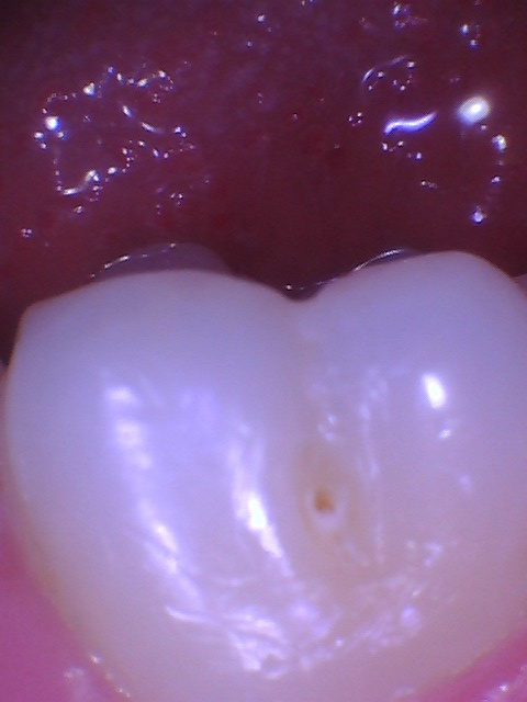
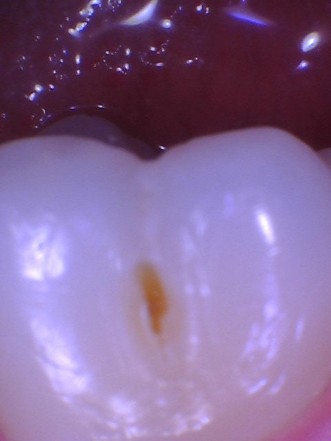
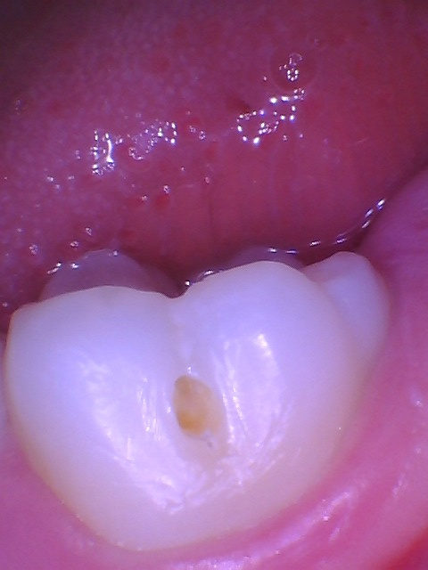
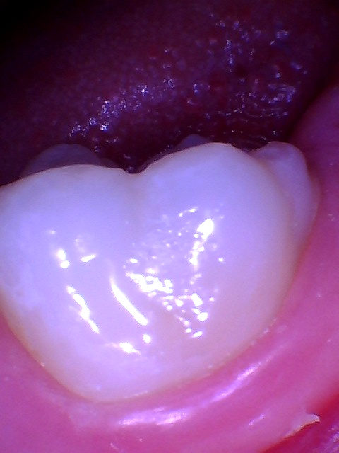
A small camera takes photographs inside your mouth and puts them on the screen next to you. When you can see the crack, or the filling that is leaking, or the gum that is inflamed, you are not deciding whether to trust a stranger. You are looking at the same picture we are.
From StartTo Finish
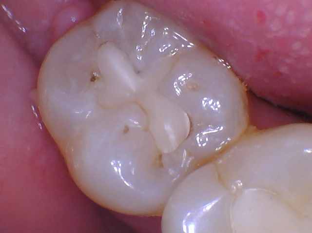
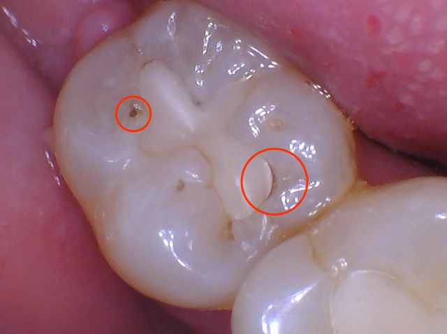
The small pit which has a cavity is on the cheek side of the tooth. Then the decay, opened. Then a tooth-colored filling, same visit.
The intraoral camera lets you see this before you decide.
AI AssistedReview
Our diagnostic software includes artificial intelligence that reviews your radiographs alongside Dr. Cortes and flags areas worth a closer look. Think of it as a second set of eyes on the image. Every diagnosis is still made by Dr. Cortes. The software does not replace his judgment, it just makes sure nothing goes unexamined.
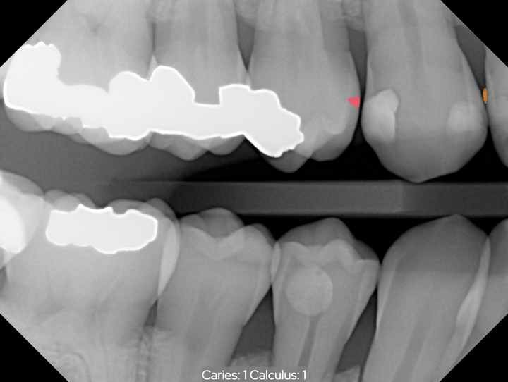
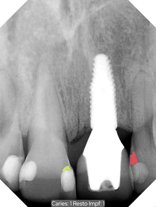
A flag is a prompt to look closer. It is not a diagnosis. Dr. Cortes reviews every one and decides what, if anything, it means.
Why it matters
The DecisionIs Yours
None of this is really about equipment. It is about who gets to make the decision.
Dentistry has traditionally worked one way. The dentist looked, the dentist decided, and the patient was told. You were asked to agree to something you could not see, on the judgment of someone you had often just met.
Putting the image on a screen in front of you changes that completely. You see the crack. You see the shadow under the old filling. You see where the bone sits. And once you can see it, nobody is persuading anybody. We are looking at the same evidence and working out what to do about it together.
That is what the technology is actually for. Not to impress you, and not to speed us up. It is to give you enough information to make the right call for your own health, which is the only person's call it ever should have been.